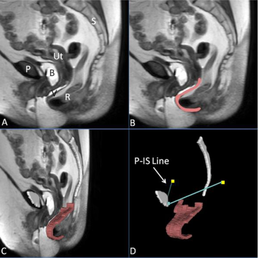
Fig 1. Making a 3D prolapse model including the P-IS line.
(A) Mid-sagittal MR image of subject with posterior prolapse; (B) Outline of posterior vaginal wall in pink; (C) Addition of midsagittal pelvic bones (white) and 3D model of posterior vaginal wall shown in slightly skewed sagittal image; (D) Straining posterior vaginal wall model and its relationship to the normalized ATFP, shown here as the turquoise lines extending from the public symphysis to the ischial spines (yellow squares), or the P-IS line. P, pubic symphysis; S, sacrum; B, bladder; R, rectum; V, vagina; Ut, uterus; IS, ischial spine. (© DeLancey 2011)

