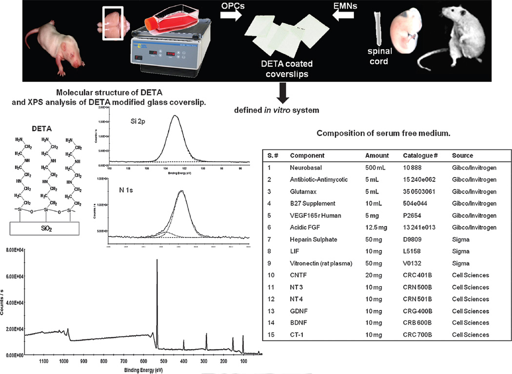
Fig. 1.
Schematic representation of co-cultures of OPCs with EMNs in defined in vitro system. Diagram illustrates the main steps in the OPCs–EMNs co-culture and development of a defined in vitro system. This system involves modification of a glass coverslip with a monolayer of the synthetic, growth promoting substrate DETA. Cells attached to DETA were cultured in the serum free medium shown, containing factors that are optimal for cell survival and the differentiation of OPCs.

