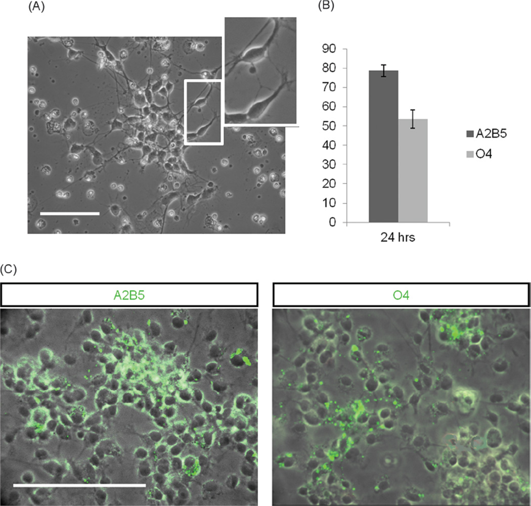
Fig. 2.
Characterization of newly isolated OPCs from the rat pup cortex. (A) A typical bipolar morphology of OPCs 24 hrs after “shake-off” isolation. (B) Results of immunocytochemical analysis revealed that 78.6±3.1% of cells expressed A2B5 and 53.6±4.7% of cells expressed O4. (C) Immunocytochemistry for A2B5 and O4 expression 24 hrs after shake off. Scale bars 100 µm.

