
Fig. 1.
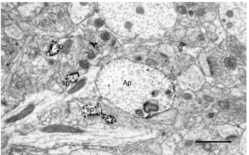
A transversely sectioned, gold toned, Golgi impregnated apical dendrite (Ap) of a layer 5 pyramidal cell. The small gold particles mark both the main shaft of the apical dendrite as well as the spines (sp) emanating from it. Scale bar = 1μm.
Official websites use .gov
A
.gov website belongs to an official
government organization in the United States.
Secure .gov websites use HTTPS
A lock (
) or https:// means you've safely
connected to the .gov website. Share sensitive
information only on official, secure websites.
A transversely sectioned, gold toned, Golgi impregnated apical dendrite (Ap) of a layer 5 pyramidal cell. The small gold particles mark both the main shaft of the apical dendrite as well as the spines (sp) emanating from it. Scale bar = 1μm.