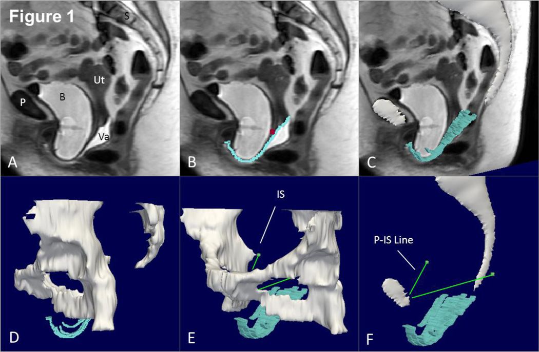
Fig 1. Making a 3D model with P-IS line.
(A) Mid-sagittal MR image of subject with prolapse. (B) Outline of anterior vaginal wall in blue with cervicovaginal junction marked with a purple square. (C) 3D model of anterior vaginal wall shown in slightly skewed sagittal image. Mid-sagittal pelvic bones also in this image. (D) Illustrates more complete view of pelvic bones and rotating this slightly in (E) we can see the ischial spine (IS). A line from the insertion of the arcus tendineus fascia pelvis on the pubic bone to the ipsilateral ischial spine is then constructed (P-IS line in E). This serves as the reference line to generate the sidewall measurements. P – pubic symphysis, S – sacrum, B- bladder, Va – vagina, Ut – uterus, IS – ischial spine. © DeLancey 2010

