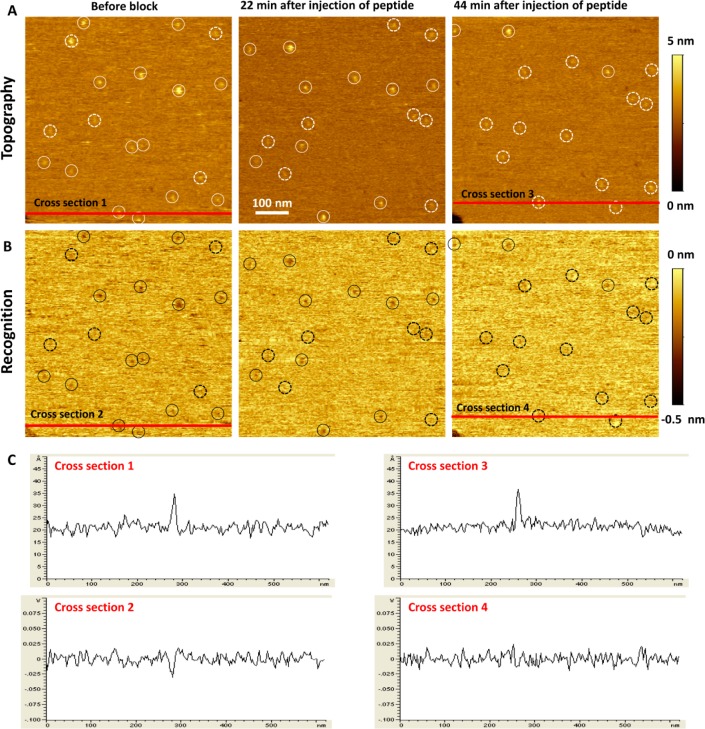
Figure 2.
High-resolution topographical (A) and UCP1 antibody-recognition (B) images of UCP1 reconstituted into a bilayer membrane. Solid and dashed circles indicate recognized and unrecognized protein molecules, respectively. Before blocking, 14 proteins are recognized and 5 proteins are not. After 44 min, nearly all molecules are blocked. (C) Cross-section images before (1,2) and after (3,4) blocking.

