
FIGURE 4. JunD influences c-Jun phosphorylation following liver I/R in a JNK1-dependent manner.
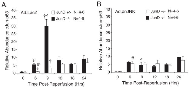
JunD−/− and JunD+/− mice were infected (intravenously) with Ad.LacZ (A) or Ad.dnJNK1 (B) viruses 3 days prior to I/R injury (partial lobar ischemia for 45 min followed by the indicated times of reperfusion). Hepatic lysates were generated from the ischemic lobe of the liver, and c-Jun was immunoprecipitated using a c-Jun antibody. Samples were then evaluated by Western blotting using a phosphoserine 63 c-Jun-specific antibody. Western blots were quantified using infrared dye-conjugated secondary antibody on a LI-COR Biosciences Odyssey Infrared Imaging System. Results depict the mean (±S.E.) for n = 4 – 6 animals at each experimental point. The 0-h time point represents non-I/R-injured control animals. *, †, #, and [caret] depict significant differences using the Student’s t test (*, †, and [caret], p < 0.01; #, p < 0.05).