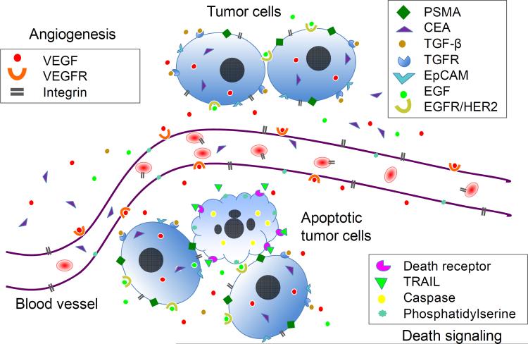
Fig. 1.
Schematic representation of potential cancer biomarkers for immuno-PET imaging. Cancer biomarkers play important roles in cancer cell proliferation, survival, angiogenesis, invasion and metastasis. The biomarkers can be associated with tumor cells (cytoplasm or surface), in the extracellular matrix (ECM) of tumor tissue, or on tumor vasculature (angiogenesis). Growth factor VEGF can be produced by tumor cells, secreted to ECM, and then bind to its receptors on endothelial cells leading to the initiation of angiogenesis. When PET imaging is employed in monitoring therapeutic efficacy, the biomarkers for cell apoptosis and necrosis, e.g. death receptors or caspase signaling can be targeted in addition to other tumor-specific targets. Note: this is for illustration purposes and does not imply that all biomarkers are present in one tumor tissue. PSMA: prostate-specific membrane antigen; CEA: carcinoembryonic antigen; TGF-β: transforming growth factor beta; TGFR: transforming growth factor receptor; EpCAM: epithelial cell adhesion molecule; EGF: epidermal growth factor; EGFR/HER-2: epidermal growth factor receptor; VEGF: vascular endothelial growth factor; VEGFR: vascular endothelial growth factor receptor; TRAIL: TNF-related apoptosis-inducing ligand.

