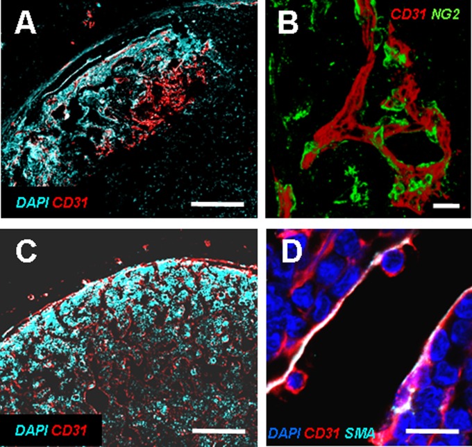
Fig. 4.
In vivo vascularization of the implanted hypertrophic cartilage templates. (A) After 5 wk only the outer region of the engineered tissue was vascularized, as shown by CD31 staining. (B) Vessels were already stabilized by NG2+ pericytes. (Scale bars, 100 µm.) (C) After 12 wk tissues were deeply vascularized, and the BM displayed sinusoid-like vascular structures with a partially SMA-positive wall, indicative of mature stabilization (D). (Scale bars, 20 µm.)

