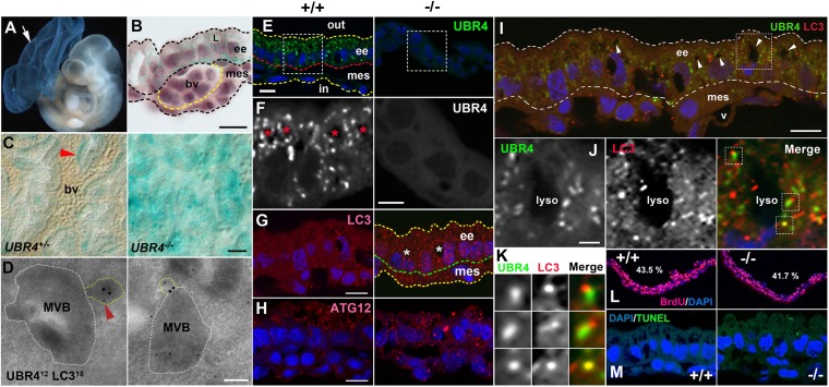
Fig. 2.
Expression of UBR4 in endoderm-derived, autophagy-enriched YS cells and its association with a constitutive autophagic pathway of the YS endoderm. (A) X-gal staining of E9.5 UBR4−/− embryo and YS. (B) Cross-section of X-gal–stained UBR4+/− YS. L, lysosome; ee, extraembryonic endoderm; mes, mesodermal layer; bv, blood vessel. (Scale bar, 10 μm.) (C) Whole-mount X-gal staining of UBR4+/− and UBR4−/− YS. Arrowhead, a relatively intense β-gal signal surrounding a blood vessel. (Scale bar, 25 μm.) (D) EM of HEK293–UBR4V5 cells labeled for UBR4 (12 nm; arrowhead) and LC3 (18 nm). (Scale bar, 200 nm.) (E) Immunostaining of UBR4 on cross-sections of +/+ and UBR4−/− YS at E9.5. (Scale bar, 10 μm.) (F) Enlarged views of areas indicated by rectangles in E. (Scale bar, 5 μm.) (G and H) Cross-sections of +/+ and UBR4−/− YS stained for LC3 (G) or ATG12 (H). (Scale bar, 10 μm.) *, lysosome. (I) Cross-sections of the YS costained for UBR4 and LC3. Arrowhead, lysosome; v, blood vessel. (Scale bar, 10 μm.) (J) Enlarged views of area indicated by rectangles in I. (Scale bar, 2 μm.) (K) Enlarged views of areas indicated by rectangles in J. (L) BrdU incorporation assay of control and UBR4−/− embryos at E9.5. S-phase indices were determined to be 43.5% and 41.7% for +/+ and UBR4−/− YS, respectively. (M) TUNEL assay of +/+ and UBR4−/− YS at E9.5.

