
Fig. 1.
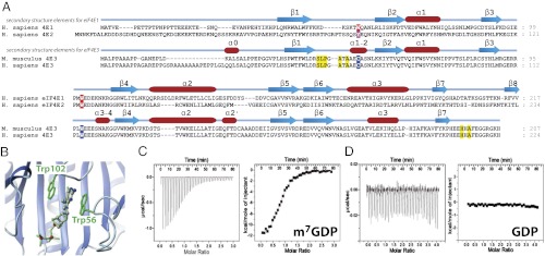
(A) Sequence alignment of the three eIF4E subfamilies. See Fig. S1 for a more comprehensive list. Secondary structures are shown. Aromatic residues important for cap recognition in eIF4E1 and eIF4E2 are highlighted in red, and corresponding residues in eIF4E3 are in blue. Other residues important for cap recognition in eIF4E3 are highlighted in yellow. (B) eIF4E1 structure (PDB ID code 3AM7) highlighting the stacking of the two aromatic residues against the m7G cap. (C and D) ITC data of wild-type eIF4E3 with m7GDP (C) and GDP (D) (Left, raw data; Right, binding isotherm).