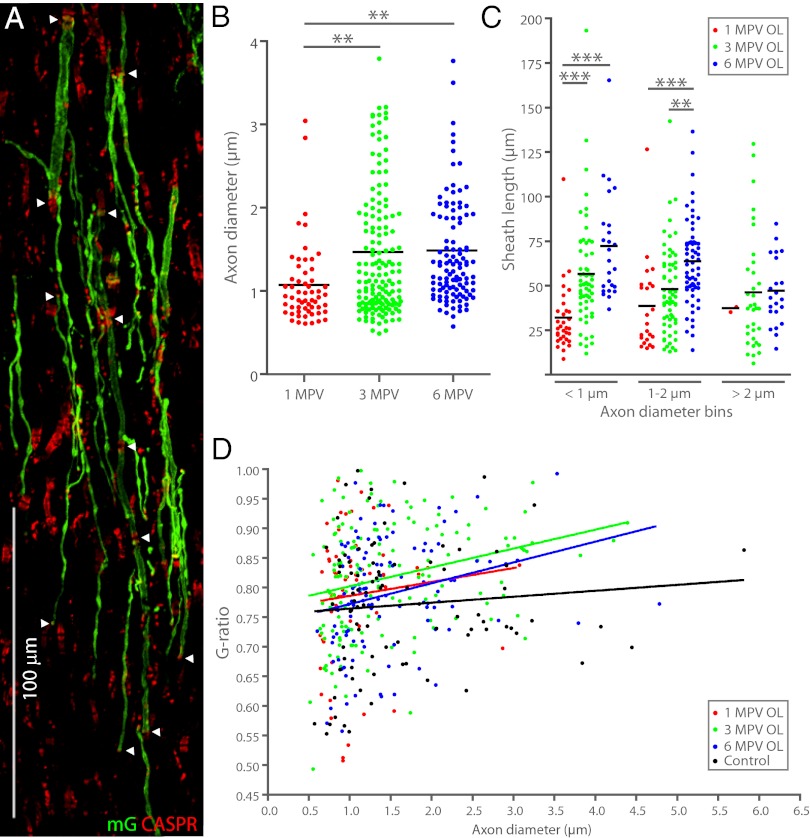
Fig. 4.
OL-regenerated sheaths exhibit postinjury remodeling. (A) OL-regenerated sheaths at 3 MPV. Arrowheads indicate CASPR+ paranodes. (B) Average axon diameter ensheathed by regenerated OLs is significantly larger at 3 and 6 MPV than at 1 MPV. (C) On small and medium-caliber axons, OL-regenerated sheaths increase in length over time. Small-caliber axons develop longer myelin sheaths than large-caliber axons, reversing the normal relationship between axon diameter and internodal length. Long, thin sheaths and short, thick sheaths can be seen in A. (D) G-ratios of regenerated and uninjured control myelin overlap considerably at all time points following injury (**P < 0.01, ***P < 0.001).

