Fig. 2.
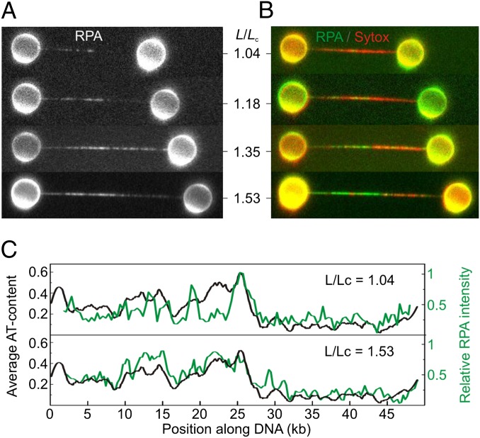
At low ionic strength (50 mM NaCl), melting bubbles nucleate in AT-rich regions. (A) Selection of fluorescence images of EGFP-RPA binding to topologically closed λ-DNA molecules as a function of relative DNA extension (L/Lc). A different DNA molecule was used for each extension. (B) Composite fluorescence images displaying the binding of EGFP-RPA (in green) and Sytox (in red). DNA molecules shown here are the same as those in A. (C) Variation in fluorescence intensity of EGFP-RPA along the DNA molecule (in green) for L/Lc = 1.04 and 1.53, respectively. Black traces display the AT-content of λ-DNA (in 100-bp bins) and are oriented in the direction that best matches the RPA fluorescence intensity (SI Text, Correlating Melting-Bubble Formation with AT-Rich DNA). These, and 10 further examples, are also shown in Fig. S5.

