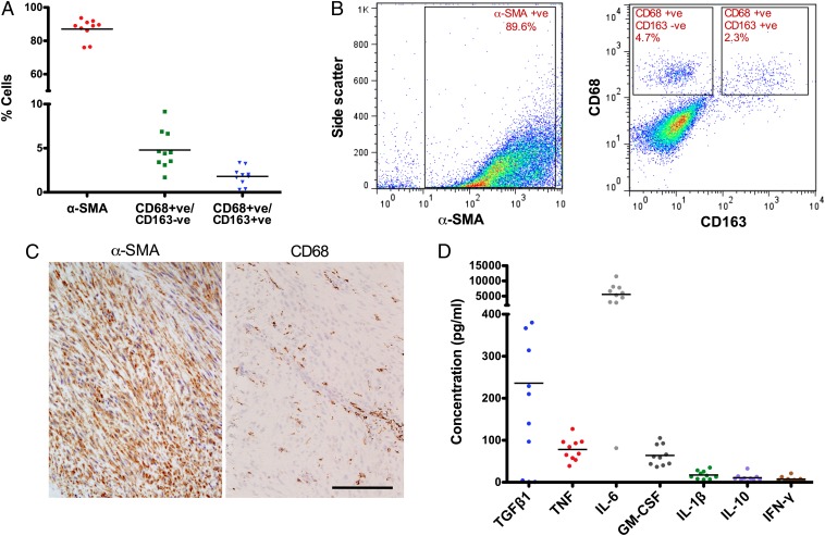
Fig. 1.
Inflammatory cells are present in Dupuytren's myofibroblast-rich nodules and resident cells in the nodules produce proinflammatory cytokines. (A) Flow cytometric analysis of cells isolated from freshly disaggregated Dupuytren's nodular tissue. Intracellular α-SMA–positive (myofibroblasts; mean ± SD: 87 ± 6.1%), cell surface CD68-positive CD163-negative (classically activated M1 macrophages; mean ± SD: 4.8 ± 2.2%), and CD68-positive CD163-positive (alternatively activated M2 macrophages; mean ± SD: 1.8 ± 1.0%) cells were quantified. (B) Representative flow cytometry scatter plots showing the proportion of α-SMA–positive cells and gating strategy for CD68+ and CD163+ cells in disaggregated Dupuytren's nodular tissue. (C) Serial histological sections of Dupuytren's nodular tissue stained for α-SMA+ (myofibroblasts) and CD68+ (monocytes) cells. CD68+ cells localized around blood vessels. (Scale bar, 100 μm.) (D) Cytokines released by freshly isolated nodular cells in monolayer culture using electrochemiluminescence. All data shown are from >10 patient samples.

