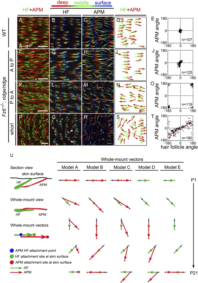
Fig. 3.
APM orientations on the back skin of P21 WT and Fz6−/−;ridge/ridge mice. (A–T) Follicles and APMs are visualized by flat mount immunostaining for GFP (Alexa Fluor 647, pseudocolored green) and smooth muscle actin (SMA; Alexa Fluor 594, red), respectively. All mice used in this experiment carry a K17-GFP transgene. Follicles and APMs are shown in Z-stacked confocal images from WT skin (A) and from different regions within a whorl in Fz6−/−;ridge/ridge skin (F, K, and P). Anterior is to the left and posterior is to the right. P to A (posterior to anterior) and A to P (anterior to posterior) refer to the global orientations of follicles in the regions shown. For each Z-stacked image, follicles (B, G, L, and Q) and APMs (C, H, M, and R) are also shown separately, with deep, middle, and surface layers pseudocolored red, green, and blue, respectively, to more clearly visualize the trajectories of individual fibers. Vector maps (D, I, N, and S) from these images show the orientations of paired hair follicles (green) and APMs (red). Scatter plots (E, J, O, and T) show the relationship between the orientations of hair follicles and their associated APMs for skin territories in and near the images shown to the left. In this and all following figures, an angle of 0° corresponds to the anterior-to-posterior direction. (Scale bar, 200 µm.) (T) Red line, Deming regression line. (U) Models of follicle (green) and APM (red) reorientation on the back skin, shown as a time series from P1 (top) to P21 (bottom) for a follicle that reorients 180°. The models differ with respect to the location, mobility, and time course of development of the APM attachment site (APMAS) at the epithelial surface. Model A: the APMAS is immobile and is located posterior to the site of follicle emergence at the skin surface. Model B: the APMAS rotates so that it always lies along the follicle axis and at the same distance from the follicle. Model C: as in model B, but APMAS rotation lags behind follicle rotation. Model D: the start of APMAS development is delayed relative to the onset of follicle rotation and the APMAS is fixed at a position that is defined by the follicle axis at an intermediate time point in development. Model E: the choice of APMAS occurs after follicle rotation has ceased and the APMAS lies along the follicle axis.

