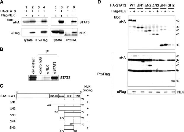
Figure 2.
STAT3 associates with NLK via its SH2 domain. (A) COS7 cells were transfected as indicated. Immunoprecipitates obtained by using anti-Flag (lanes 3,4) or anti-HA (lanes 7,8) antibody were subjected to Western blotting with the indicated antibodies. Expression of HA-STAT3 or Flag-NLK was monitored (lanes 1,2,5,6). (B) Immunoprecipitates obtained from mouse embryonic fibroblast extracts by using control IgG, anti-NLK antibody, or anti-STAT3 antibody were subjected to Western blotting with anti-STAT3 antibody. (C) Diagram of STAT3 deletion mutants. (D) 293 cells were transfected as indicated. Immunoprecipitates obtained with anti-Flag antibody were subjected to Western blotting with anti-HA antibody (middle) or anti-Flag antibody (bottom). Expression of HA-STAT3 deletion mutants was monitored (top). Open triangles and closed triangles indicate the migrations of STAT3 deletion mutants and IgG proteins, respectively.

