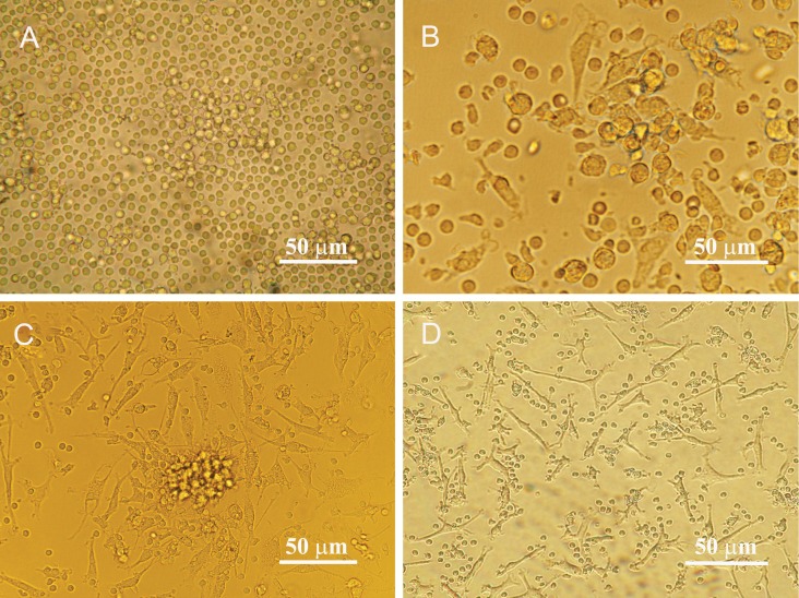
Fig 1.
Morphological features of isolated endothelial progenitor cells before and after differentiation. A. Mononuclear cells isolated from human peripheral blood appeared small and round. B. Most cells were attached to the plates coated with human fibronectin (day 4). The attached cells gradually exhibited a spindle-shaped, endothelial cell-like morphology on day 7th C. and day14th D. after seeding.

