Fig 3.

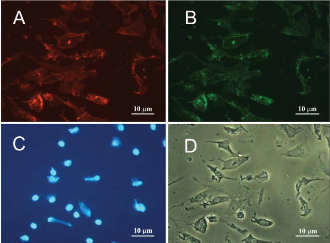
A. Immunocytochemistry of EPCs double-stained with anti-CD31, and B. anti-CD34 antibodies. C. Cells' nuclei were visualized with DAPI . D. The same phase contrast view of the cells in A-C.

A. Immunocytochemistry of EPCs double-stained with anti-CD31, and B. anti-CD34 antibodies. C. Cells' nuclei were visualized with DAPI . D. The same phase contrast view of the cells in A-C.