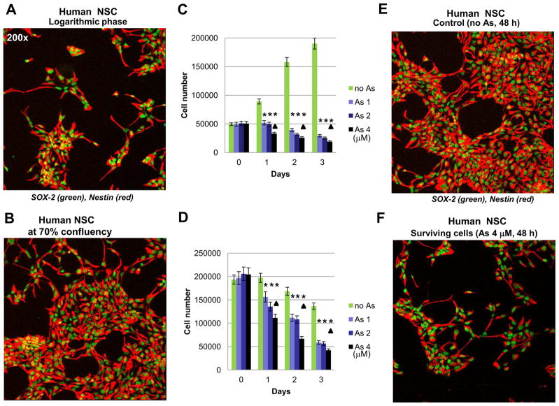
Figure 1.
Effects of sodium arsenite treatment on growth and survival of human neural stem cells (NSC). (A, B, E and F) Confocal analysis of immunofluorescent images of NSC was performed using monoclonal antibody against Nestin, an early neuroprogenitor cell marker (red), and polyclonal antibody against Sox2, a pluripotency marker (green). (C and D) Determination of human NSC growth and survival was performed in the absence or in the presence of increased doses of sodium arsenite (1–4 μM). Sodium arsenite was added to NSC cultures at the logarithmic phase (panels A and C) or at 70% confluency (panels B and D) at day 0 for the next 3 days. Numbers of live cells attached to fibronectin matrix were determined. Pooled results of five independent experiments are shown in panels C and D. Error bars represent means ± SD. Statistically significant differences in cell number between control and arsenite treated (1–4 μM) cell cultures are indicated by the stars. Statistically significant differences in cell survival between cell cultures treated with 4 μM arsenite, compared to cultures treated with 1 μM arsenite, are indicated by the triangles (p<0.05, Student’s t test). Images of control NSC and surviving NSC 48 h after sodium arsenite exposure are shown on panels E and F.

