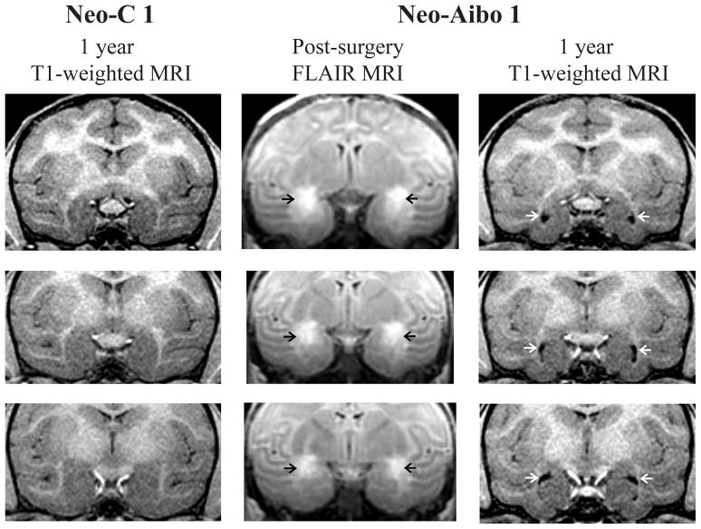
Figure 1.
Coronal sections through the anterior (top) to posterior (bottom) extent of the amygdala. Left column: T1-weighted MR images through the amygdala at 1-year post-surgery in one sham-operated control (Neo-C-1). Middle column: FLAIR images illustrating the location and extent of hypersignals (black arrows) within the amygdala in a representative case with neonatal amygdala lesion (Neo-Aibo-1). Rigtht column: T1-weighted MR images through the amygdala at 1-year post-surgery in case Neo-Aibo-1 illustrating the enlargement of ventricles (white arrows) resulting from amygdala volume reduction.

