Figure 1.
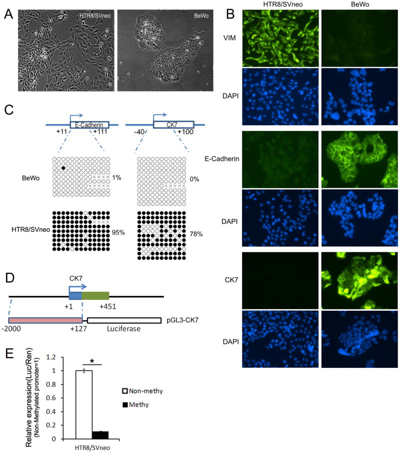
Differences in morphology, protein expression and DNA methylation status between BeWo and HTR8/SVneo cells suggest that the differentiation process from cytotrophoblast (CT) to extravillous trophoblast (ECT) is accompanied by Epithelial-to-Mesenchymal transition (EMT). A, The morphology of HTR8/SVneo and BeWo cells. B, Immunocytochemical analysis of E-Cadherin, VIM and CK7 in these cells. Original picture is 200X. C, DNA methylation of proximal promoter of E-Cadherin and CK7 gene. The CpG sites from +11 to +111 bps (relative to transcription start site) of E-Cadherin gene and from −40 to +100 bps of CK7 gene were included in bisulfite genomic sequencing assay respectively. The top panel showed genomic regions of E-Cadherin and CK7 genes. DNA methylation data was analyzed by quantification tool for methylation analysis online software (http://quma.cdb.riken.jp/). DNA methylation of CpG sites were represented by cycles, with open cycles and close cycles describing unmethylated and methylated CpG sites respectively, and the “x” decoding non-sequenced CpG sites. D, Schematic diagram of pGL3-CK7 construct used in luciferase reporter assay. E, Effect of in vitro DNA methylation of CK7 promoter activity in HTR8/SVneo cells. Data is presented as mean ± SEM.

