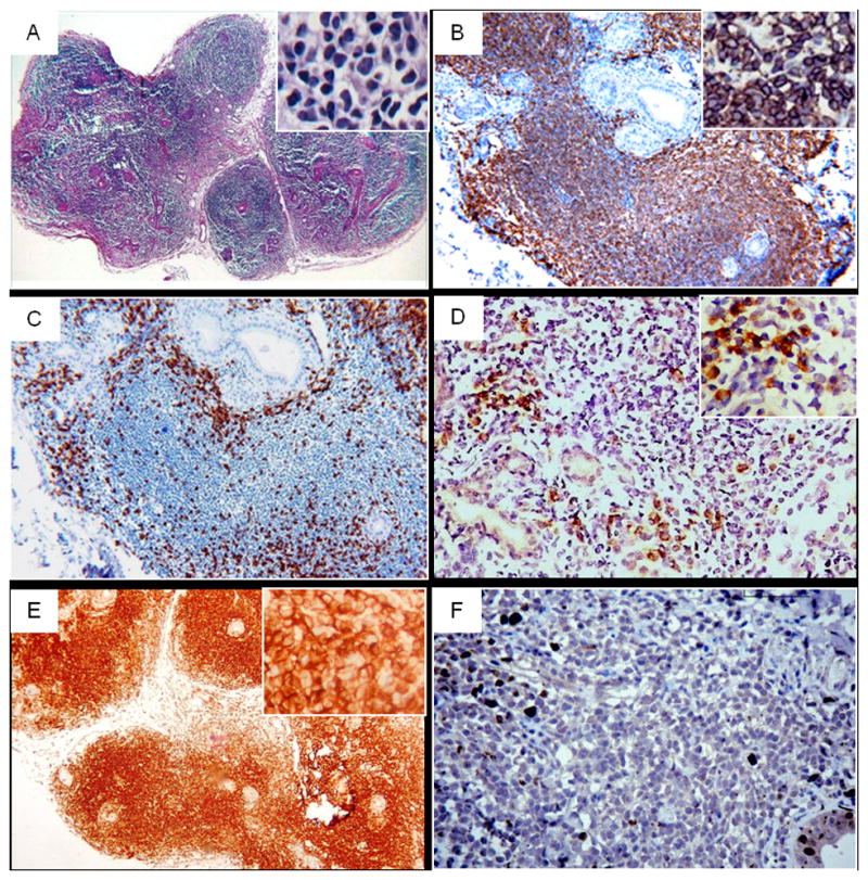
Fig. 1.

Histomorphology and immunohistochemistry of MALT lymphoma in minor salivary gland follow up biopsy; A: Hematoxylin-eosin (Original magnification ×40), B: CD20 (positive) in most lymphocytes exhibiting a diffuse pattern, C: CD3 (negative) identifies scattered T-cells of the lymphocytic infiltrate, D: Kappa light chain restriction, E: Bcl-2 positive staining, F: Ki-67 positive expression <5% of neoplastic cells. (Original magnification ×100).
