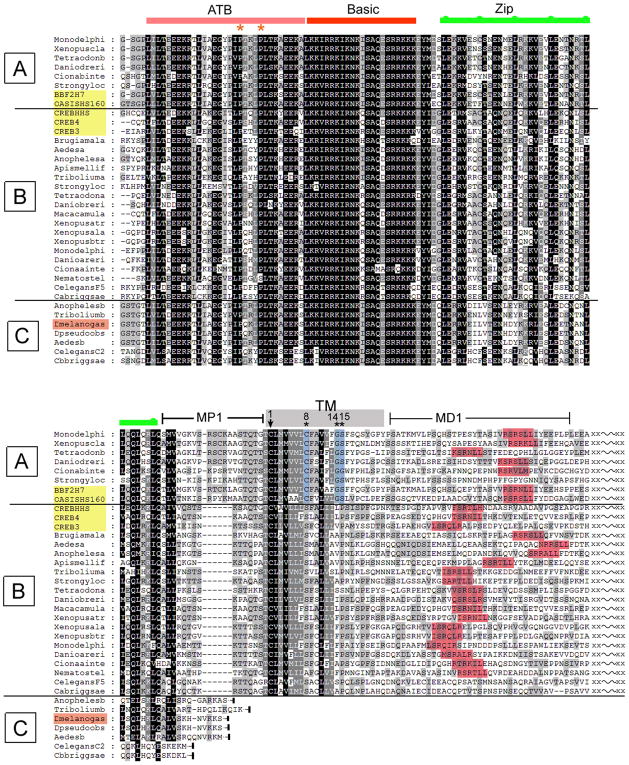
Figure 7. Evolutionary conservation of the CREB3 family and identification of distinct classes.
The figure shows only representative proteins at the amino acid level to allow for key points regarding subgrouping and evolutionary history to be made. For example, numerous CREB3 species from mammals have been omitted, but these all fit with the conclusions and classification discussed in the text. The family of five human CREB3 proteins are shaded in yellow, and thus resolve into the A and B classes. MP1 indicates membrane proximal region 1; TM, transmembrane; MD1, membrane distal region 1. Consensus S1P sites are shaded red. Numbering within the TM domain starts at the completely conserved cysteine. Other features are as described in the text.

