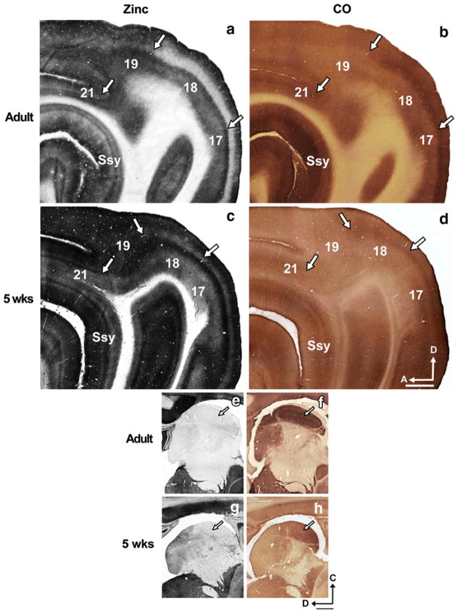
Fig. 1.
Synaptic zinc staining in the adult and juvenile ferret brain distinguishes different visual cortical areas. Photomicrographs of adjacent semi-tangential sections stained for a synaptic zinc or b cytochrome oxidase (CO) in the adult, and c synaptic zinc or d CO in the 5-week-old juvenile. Arrows mark areal boundaries. Photomicrographs of adjacent semi-tangential sections of thalamus stained for e synaptic zinc or f CO in the adult, and g synaptic zinc or h CO in the 5-week-old juvenile. Arrows indicate lateral geniculate nucleus. Ssy Suprasylvian cortex, A anterior, D dorsal. Scale bar 1 mm

