
Figure 2.
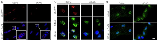
Nuclear foci of CUGexp RNA and MBNL1 protein are dispersed in muscles treated with siCAG. (a) Fluorescence in situ hybridization (FISH) shows reduction of CUGexp foci (red) in nuclei (blue) in isolated flexor digitorum brevis (FDB) muscle fibers. Muscle was taken 7 days after electroporation of siCAG or saline. Inset shows magnification of selected nuclei. Nuclei are counterstained blue using 4,6 diamino-2-phenylindole dihydrochloride (DAPI). (b) Combined FISH for CUGexp RNA (red) and immunofluorescence for MBNL1 protein (green) in transverse sections of tibialis anterior muscle from human skeletal actin—long repeat mice. Muscle was taken 7 days after electroporation of siCAG or saline. The CUGexp foci are dispersed and the MBNL1 distribution is more diffuse in siCAG-treated muscle. Nuclei are counterstained blue (DAPI). (c) Immunofluorescence of isolated FDB muscle fibers shows redistribution of MBNL1 (green) from punctate to diffuse localization in the nucleus (blue), 7 days after electroporation of siCAG.Scale bars = 5 µm. Objective: 100× Plan Apo, 1.4 NA oil.