Figure 5.

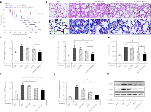
Improvements of survival and lung fibrosis by Ad-sTβR-MSCs treatment. (a) Effects of Ad-sTβR-MSCs on long-term survival (*P < 0.05, **P < 0.01, n = 10). (b) Hematoxylin and eosin (H&E) (upper panel) and Masson's trichrome staining (lower panel) of lung sections, examples of focal fibrotic lesions (arrows) are marked. Bar = 50 µm. (c) Malondialdehyde (MDA) levels, (d) hydroxyproline content in lung homogenates, and active (e) transforming growth factor-β1 (TGF-β1) concentrations in plasma (*P < 0.05, **P < 0.01, n = 5–8). (f–g) The mRNA and (h) protein levels of connective tissue growth factor (CTGF) and α-smooth muscle actin (α-SMA) in lungs were analyzed by quantitative reverse transcription-PCR (RT-PCR) and western blot assays (*P < 0.05, **P < 0.01, n = 5–8). MSCs, mesenchymal stem cells.
