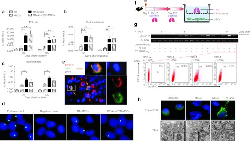
Figure 6.

Mesenchymal stem cells (MSCs) adopted alveolar type II (ATII) cells phenotype. (a–c) Quantitative real-time PCR analysis of the male MSCs rates in isolated lung (a) ATII cells, (b) endothelial cells, and (c) myofibroblasts from mice receiving radiation therapy (RT), MSCs, RT+MSCs, or RT+Ad-sTβR-MSCs (*P < 0.05, **P < 0.01, n = 5). (d) Y chromosome FISH assay in ATII cells from male mice (positive control), control female mice (negative control) and female mice of RT+MSCs or RT+Ad-sTβR-MSCs group, magnification ×1,000. (e) Immunofluorescence (IF) of frozen lung sections from RT+Ad-sTβR-MSCs group on days 30 by using de-convolution microscopy. Nuclear staining (DAPI, blue), Ad-sTβR-MSCs (GFP, green), and the ATII cells (proSP-C, red), magnification ×400. (f) Schematic representation of coculture assay ex vivo. MSCs were cocultured with irradiated lung tissue. (g) The proSP-C mRNA expression was detected by RT-PCR in MSCs cocultured with the uninjured lung, RT 7d lung and RT 120d lung (upper panel). The proSP-C positive cells were counted by fluorescence-activated cell sorting (FACS) 14 days after coculture (lower panel). (h) The proSP-C expression and lamellar bodies in MSCs cocultured with RT 7d lung for 14 days were detected by IF (upper panel) and transmission electron microscopy (TEM) (lower panel), respectively. IF, magnification ×1,000. TEM, magnification ×12,000.
