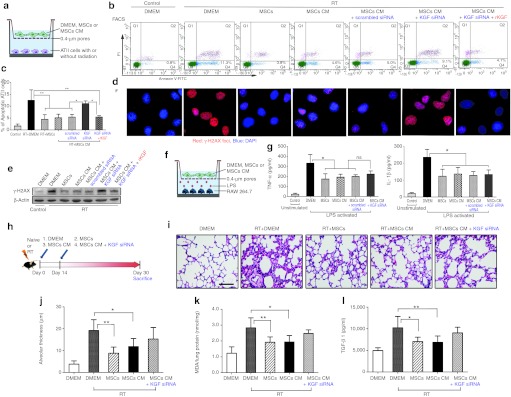
Figure 7.

Protective effects of mesenchymal stem cells (MSCs)-conditioned medium (CM) in vitro and in vivo. (a) Schematic representation of in vitro incubation/coculture assay of radiation-injured alveolar type II (ATII) cells with DMEM, MSCs, or MSCs-conditioned medium (MSCs CM). (b) Fluorescence-activated cell sorting (FACS) analysis of the apoptotic irradiated-ATII cells, when incubated/cocultured for 48 hours with DMEM, MSCs, MSCs CM, MSCs CM in the presence of keratinocyte growth factor (KGF) siRNA or plus recombinant KGF (rKGF). (c) The statistic results of FACS (*P < 0.05, **P < 0.01, n = 5). (d) The DNA damage was assayed by γ-H2AX staining for irradiated ATII cells incubated/cocultured for 24 hours with DMEM, MSCs, MSCs CM, MSCs CM in the presence of KGF siRNA or plus rKGF. Magnification ×1,000. (e) Western blot analysis of γ-H2AX. (f) Schematic representation of in vitro incubation/coculture assay of lipopolysaccharide (LPS)-stimulated macrophage RAW264.7 with of DMEM, MSCs, or MSCs CM. (g) Enzyme-linked immunosorbent assay (ELISA) analysis of the tumor necrosis factor-α (TNF-α) and IL-1β concentrations in the medium of LPS-activated RAW264.7 incubated/cocultured for 24 hours with DMEM, MSCs, MSCs CM, or KGF siRNA-pretreated MSCs CM (*P < 0.05, **P < 0.01, n = 5). (h) Schematic representation of in vivo effects of MSCs, MSCs CM, or KGF siRNA-pretreated MSCs CM on radiation-induced lung injury (RILI) 30 days after irradiation. (i) Representative hematoxylin and eosin (H&E) stained lung sections from five experimental groups. Bar = 50 µm. (j) The radiation-induced lung damage was semiquantified by measuring the alveolar thickness (*P < 0.05, **P < 0.01, n = 5). (k) Malondialdehyde (MDA) levels in the lung homogenates were assayed (*P < 0.05, **P < 0.01, n = 5). (l) Enzyme-linked immunosorbent assay (ELISA) analysis of the plasma active transforming growth factor-β1 (TGF-β1) (*P < 0.05, **P < 0.01, n = 5).
