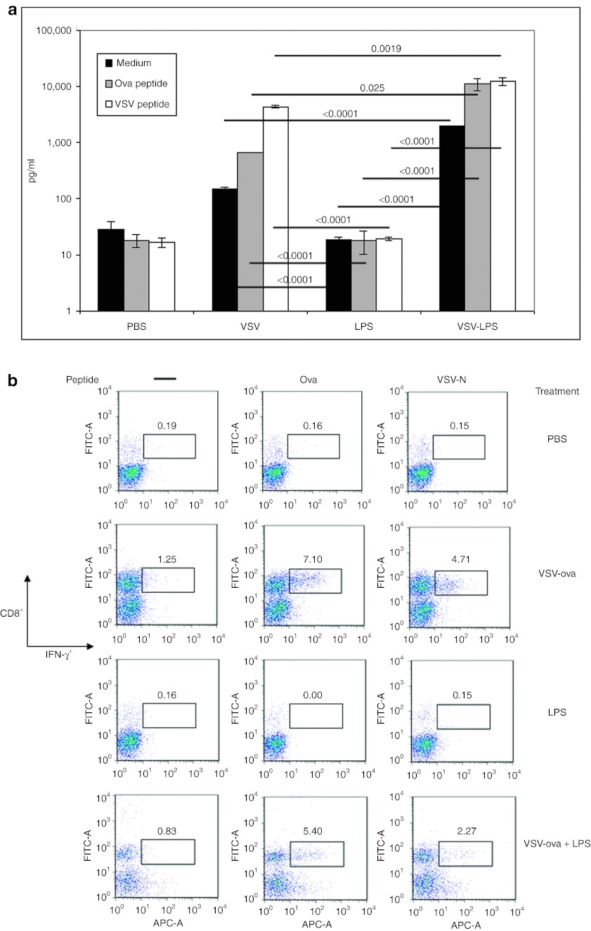
Figure 3.
Shift in generalized and antigen-specific T-cell activation in mice treated with VSV-ova + LPS. (a,b) C57BL/6 mice bearing 7-day subcutaneous tumors (n = 8/group) were administered intratumorally VSV-ova (5 × 10⁸ PFU/50 µl), PBS (50 µl), and/or LPS (200 µg/50 µl). LPS (200 µg/50 µl), when given in combination with VSV-ova, was given on day 8. (a) Tumor-draining lymph nodes and (b) tumors were harvested on day 14 (n = 3/group). Samples were pulsed with either no peptide (medium), ova or VSV-specific peptides for 48 hours (in a) or 1 hour (in b) with T-cell activation being assessed by IFN-γ ELISA of cell-free supernates (in a) or intracellular IFN-γ staining (in b). Data are shown as mean ± SD where appropriate. ELISA, enzyme-linked immunosorbent assay; IFN, interferon; LPS, lipopolysaccharide; PBS, phosphate-buffered saline; ova, ovalbumin; PFU, plaque-forming unit; VSV, vesicular stomatitis virus.

