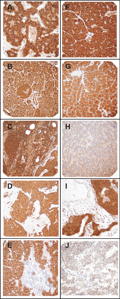
Figure 1.

Immunohistochemical analysis of pancreatic endocrine tumors. Panels illustrate individual PET specimens in the TMA (original magnification × 200) exhibiting strong immunostaining for (A) VEGFR1, (B) TGFBR1, (C) PDGFRA, (D) SSTR5, (E) SSTR2A, (F) IGF1R, (G) Hsp90, (H) EGFR, (I) mTOR, and (J) MGMT.
