Figure 3.
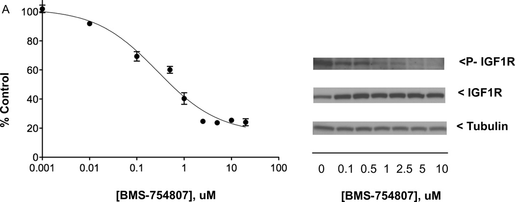
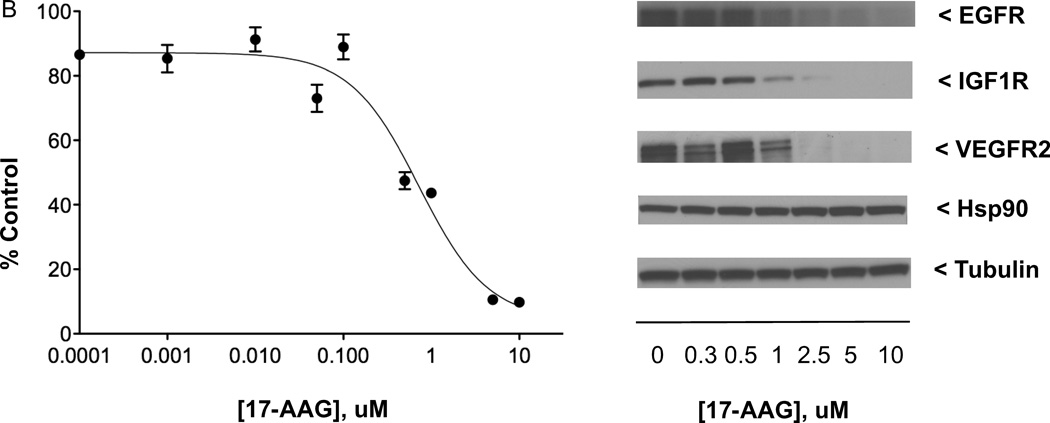
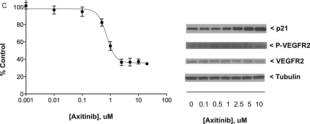
Effects of anticancer drugs in QGP-1 PET cell line. (A) Left: QGP-1 cells (4000/well) were incubated (continuous exposure, 5 days) in 96-well plates at 37°C with increasing concentrations of anticancer drug BMS-754807 (targeting IGF1R/IR) in serum-containing medium, with cell proliferation measured by the MTS assay; right: QGP-1 cells (500,000/well) were incubated (continuous exposure, 24 h) in 6-well plates at 37°C with increasing concentrations of BMS-754807 in serum-containing medium, with effect on constitutive IGF1R phosphorylation determined by western immunoblotting of equal quantities of protein from whole cell lysates. (B) Effect of increasing concentrations of anticancer drug 17-AAG (targeting Hsp90) on QGP-1 cell growth and levels of indicated biomarkers, with experiments performed as described in (A). (C) Effect of increasing concentrations of anticancer therapeutic axitinib (targeting VEGFR1–3, PDGFRA-B, and KIT) on QGP-1 cell growth and levels of indicated biomarkers, with experiments performed as described in (A). MTS data presented were the average ± SE of three experiments; western immunoblotting results were representative blots from one of three experiments.


