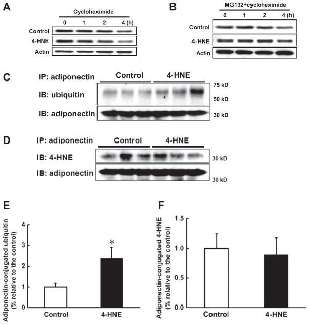
Fig. 5.
4-HNE accelerated adiponectin degradation via ubiquitin–proteasome system. After 2-h pretreatment with cycloheximide (5 μg/ml), 3T3-L1 adipocytes were exposed to 4-HNE (30 μM) for 1, 2 or 4 h and the total proteins were isolated to detect intracellular adiponectin protein abundance via Western blot. (A) 4-HNE reduced adiponectin protein stability. Proteasome inhibitor, MG132 (10 μM), was added to the medium 2 h before cycloheximide (5 μg/ml) treatment. Two more hours later, 3T3-L1 adipocytes were exposed to 4-HNE (30 μM) for 1, 2 or 4 h and intracellular adiponectin protein abundance was detected via Western blot. (B) MG132 abolished 4-HNE induced enhancement of adiponectin protein degradation. MG132 (10 μM) was added to the medium for 2 h before 4-HNE (30 μM) exposure. Four hours later, the proteins were isolated and immunoprecipitated by adiponectin antibody. The pull-down proteins were subjected to Western blot and probed with ubiquitin or 4-HNE antibodies to determine adiponectin-conjugated ubiquitin and 4-HNE-adiponecitn adduct levels. (C & E) 4-HNE increased adiponectin-conjugated ubiquitin contents in 3T3-L1 adipocytes in comparison to control cells. (D & F) 4-HNE exposure had no effects on 4-HNE-adiponectin adduct formation in 3T3-L1 adipocytes. Data are means ± SD (n = 3). *P < 0.05 compared with the untreated group.

