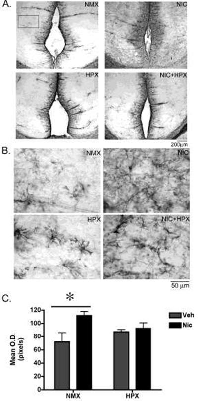
Figure 5. Prenatal nicotine but not nicotine plus hypoxia significantly increased GFAP immunoreactivity in the fetal retrosplenial (RSg) cortex.
(A,B) Representative photomicrographs of GFAP immunoreactivity (ir) in the retrosplenial cortex from fetal guinea pigs exposed in utero to nicotine (n=4), hypoxia (n=6), nicotine and hypoxia (n=5), or normoxic (n=5) conditions. The boxed field in (A) is representative of the area quantified. (B) Higher magnification photomicrographs of the RSg cortex used for quantification. (C) Quantification of GFAP-ir in the RSg cortex. Data represent bilateral optical density measurements in three sections that were averaged to yield a single value per animal. Two-way ANOVA revealed a significant main effect of nicotine (F1,19=6.6, p<0.02). Post-hoc analysis revealed that prenatal nicotine alone increased GFAP-ir in the RSg cortex compared to normoxic controls (Tukey Kramer post-hoc analysis p<0.05). All values are mean optical density in pixels ± SEM. NMX, normoxic; NIC, nicotine; HPX, hypoxic, O.D., optical density.

