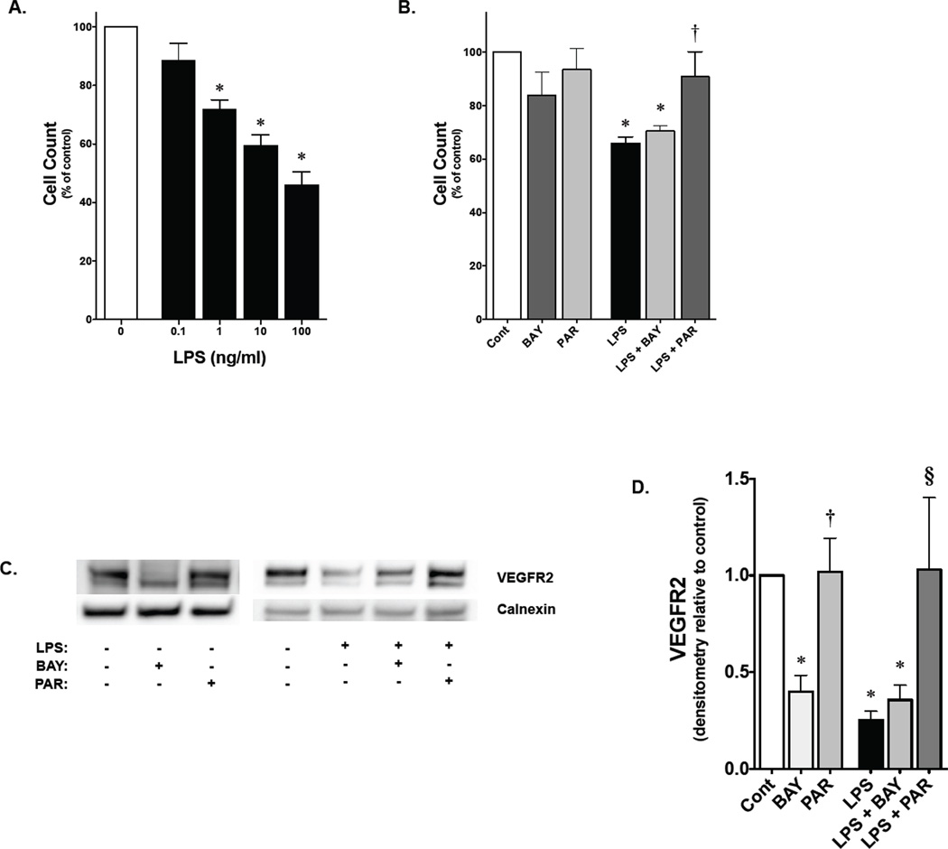
Figure 7. Parthenolide attenuates LPS-induced inhibition of fetal PAEC proliferation.
A) Fetal PAEC cell number expressed as percent of control following exposure to LPS (range, 0.1–100 ng/ml) for 3 days. *, p<0.05 vs. unexposed control. Values are means ± SE of three independent experiments for each group
B) Fetal PAEC cell number expressed as percent of control following exposure to either BAY 11-7085 (1 nmol/L), parthenolide (0.1 µmol/L), LPS (1 ng/ml) or combination for 3 days. *, p<0.05 vs. control; †, p<0.05 vs. LPS and LPS+BAY exposed. Values are means ± SE of three independent experiments for each group.
C) Representative Western blot showing VEGFR2 protein in whole cell lysates from fetal PAEC pretreated with BAY-7085 (4 µmol/L, 1h) or parthenolide (2 µmol/L, 1h) prior to LPS exposure (10 ng/mL, 6h). Calnexin is shown as a loading control.
D) Densitometric evaluation of VEGFR2 in fetal PAEC pretreated with NF-κB inhibitors (1h) and exposed to LPS (10 ng/mL, 6h). *, p<0.05 vs. control. †, p<0.05 vs. BAY-7085 pre-treatment; §, p<0.05 vs. time matched LPS and BAY+LPS exposed. Values are means ± SE of three independent experiments for each group.

