Figure 2.
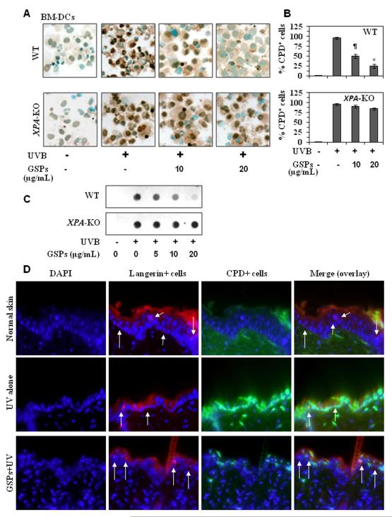
GSPs significantly repair UVB-induced DNA damage in BM-DCs obtained from wild-type mice but do not repair damaged DNA in BM-DCs from XPA-KO mice. A, BM-DCs were exposed to UVB (5 mJ/cm2) with or without the pretreatment with GSPs (0, 5, 10 and 20 μg/mL) and harvested either immediately or 24 hours later, cytospun, and subjected to cytostaining to detect CPD+ cells. CPD+ cells are dark brown. Magnification, x400. B, The numbers of UVB-induced CPD+ cells in wild-type and XPA-deficient BM-DCs are expressed in terms of the percentage of CPD+ cells as a mean ± SD of the results of three independent experiments. Significant difference vs GSPs-treated and UVB-irradiated cells, *P<0.001; ¶P<0.01. C, Analysis of CPDs by dot-blot assay. Results are shown from a single experiment that is a representative of two separate experiments. D, Treatment of mice with dietary GSPs enhanced the repair of UV-induced DNA damage in epidermal DCs (langerin-positive cells). Red fluorescence indicates langerin+ cells and green fluorescence indicates CPD+ cells. Arrows indicate langerin-positive and CPDs-negative or less intense cells in GSPs-fed group under overlay (merge) panel. Representative photomicrographs are shown, n=3/group. Magnification, x400.

