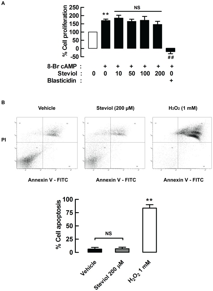
Figure 3. Effect of steviol on cell proliferation and apoptosis.
(A) MDCK cell proliferation was assessed by BrdU cell proliferation assay. MDCK cells were seeded onto 96-well plates and grown for 24 h. The media containing 8-Br cAMP (100 µM) with or without steviol at doses of 10, 50, 100, and 200 µM was added and incubated for 24 h. BrdU was added at 18 h after addition of all compounds. The data represent percent of cell proliferation of MDCK cells treated with steviol at various concentrations. 20 µg/ml of blasticidin was used as positive control. Four independent experiments were done (mean of percent control±SE; n = 4, **P<0.01 compared with group of no cAMP treatment; ## P<0.01 compared with cAMP treated group). (B) MDCK cell apoptosis was analyzed by flow cytometry. (3B, top) MDCK cell were incubated with steviol at a concentration of 200 µM for 24 h. Cells were stained with annexin V or propidium iodide. Apoptotic cells were localized in the lower right (early apoptosis) and upper right (late apoptosis) quadrants of the dot-pot graph using propidium iodide vs annexin V. 1 mM of hydrogen peroxide (H2O2) was used as positive control. (3B, bottom) The bar graphs represented percent of MDCK cell apoptosis of four independent experiments (mean percent control±SE; n = 4; **P<0.01 compared with control).

