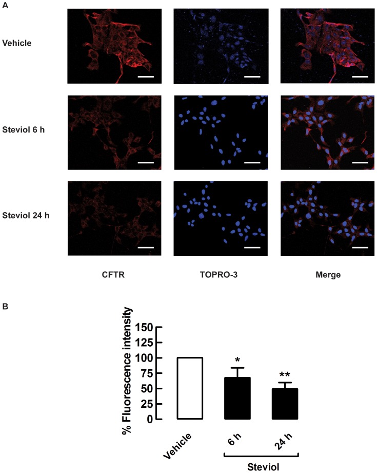
Figure 8. Steviol inhibition of CFTR membrane protein expression in MDCK cells.
(A) Representative immunofluorescence images of CFTR (red), TOPRO-3-lebeled nuclei (blue) and merged images (n = 3). Scale bar = 50 µm; magnification = ×40. (B) Mean fluorescence intensity in MDCK cell after treatment with DMSO (vehicle) or 100 µM steviol (experimental) for 6 h and 24 h. The values are shown as percent fluorescence intensity (35 random regions of interest; mean percent of control±SE, *P<0.05, **P<0.01 compared with control).

