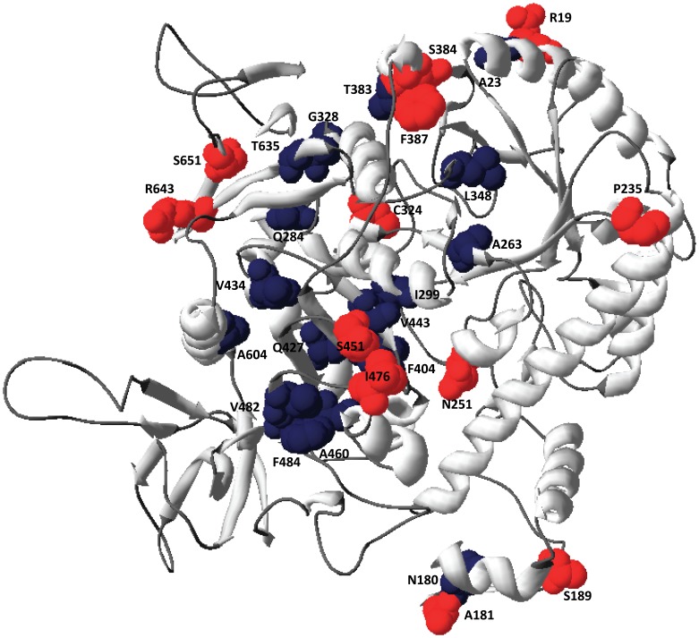
Figure 4. Model of H. lacusprofundi β-galactosidase highlighting differences with mesophilic Haloarchaea.
The protein backbone is colored gray, substitutions of surface residues are shown colored red, and substitutions of internal residues are shown colored dark blue. The protein structure was illustrated using Swiss-PDBViewer [65].

