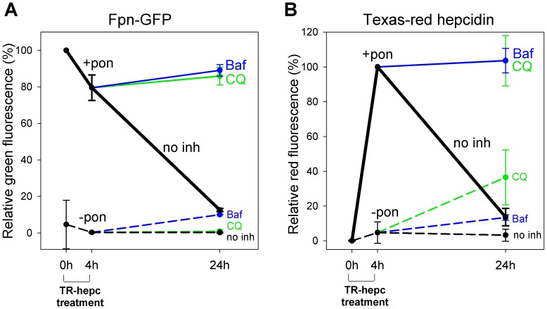
Figure 3. Hepcidin is degraded together with ferroportin.
ECR293cells were either not induced (-pon, dashed lines) or were induced with ponasterone to express Fpn-GFP (+pon, solid lines). Cells were treated with Texas Red-labeled hepcidin (TR-hepc) for 4 h, the peptide was then removed, and cells were incubated for another 20 h in the absence or presence of lysosomal inhibitors chloroquine (100 µM, green lines) or bafilomycin (100 nM, blue lines). Cells treated with hepcidin but not inhibitors are represented as black lines (no inh). The amount of cellular Fpn-GFP or Texas Red-hepcidin was determined by flow cytometry. Uninduced cells were used to establish a gate to exclude background fluorescence. (A) Quantitation of Fpn-GFP. Data are expressed relative to the green fluorescence of induced cells not treated with hepcidin. (B) Quantitation of Texas Red-hepcidin. Data are expressed relative to the red fluorescence of induced cells treated with Texas Red-hepcidin for 4 h.

