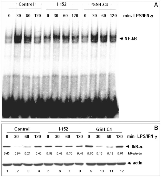
Figure 5. NF-kB activation in murine peritoneal macrophages.
(A): NF-kB nuclear translocation and DNA binding activity was assessed by Electrophoretic Mobility Shift Assay (EMSA) of nuclear extracts obtained from macrophages untreated (Control) or pre-treated with either 20 mM GSH-C4 or 20 mM I-152 for 2 hours and then stimulated with LPS and IFN-γ after molecule removal, for different times as indicated. (B): IkB-α degradation and re-synthesis as monitored by Immunoblotting analysis of IkB-α protein levels in the cytosolic fraction of macrophages treated as above. Cytosolic proteins (15 µgs) were separated by SDS-PAGE onto 10% acrylamide gels, transferred to nitrocellulose membrane and immunoblotted with an anti-IkB-α antibody. Actin was stained as a loading control. IkB-α densitometric values, normalized to actin, are reported immediately below the IkB-α blot.

