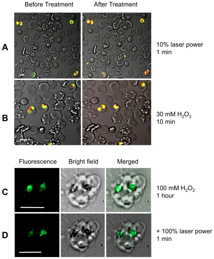
Figure 5. The parasite DV remains intact in parasites subjected to oxidative stress.
(A) and (B) are confocal micrographs showing the redistribution of acridine orange fluorescence in intact parasitized erythrocytes subjected either to (A) 1 min illumination with the microscope's laser, or (B) 10 min exposure to 30 mM H2O2. Prior to the treatment the DV fluoresces red and the parasite cytosol fluoresces green. Both treatments resulted in a loss of red fluorescence from the region of the DV. (C) and (D) show the retention of fluorescein-dextran within the DV of mature, isolated D10 trophozoites (there are two visible in the image) following exposure to 100 mM H2O2 for 1 hour (C), followed by excitation with a 488 nm laser line at full power for 1 min (D). Neither the high concentration of H2O2 alone, nor the subsequent additional intense light exposure resulted in any redistribution of the fluorescein-dextran from the DV of the parasites (visible as dark regions, coinciding with the hemozoin crystals, in the bright-field images), from which it may be concluded that the DV remained intact. All scale bars (shown in white) are 5 µm.

