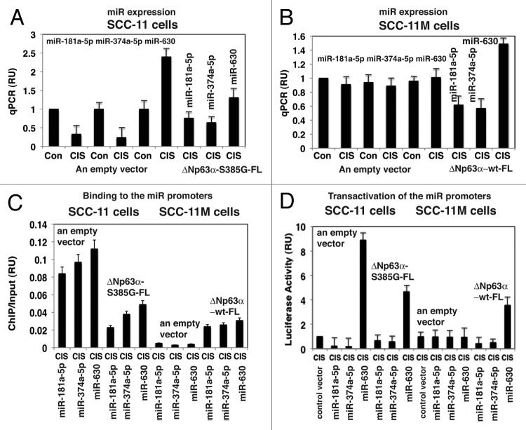
Figure 1. P-Δ;Np63α regulated transcription of the specific microRNA promoters in SCC-11 cells upon cisplatin exposure. SCC-11 cells (A) and SCC-11M (B) cells were transfected with an empty vector and with the Δ;Np63α-S385G-FL or Δ;Np63α-wt-FL expression cassettes for 24 h, exposed to control media or 10 μg/ml cisplatin (CIS) for 12 h and then tested for specific microRNA expression using qPCR (A and B) qPCR experiments were performed in triplicate with +SD as indicated (< 0.05). (C) Resulting SCC-11 and SCC-11M cells were used for ChIP analysis to identify the binding of Δ;Np63α to the specific microRNA promoters. The amount of immunoprecipitated-enriched DNA in each sample (ChIP) is represented as signal relative to the total amount of input chromatin DNA (input) using the same primers for the specific promoter region. (D) SCC-11 cells and SCC-11M cells were also transfected with 100 ng of the control promoter-less pLightSwitch_Prom plasmid and the pLightSwitch_Prom plasmids containing promoter sequences of specific microRNAs (as indicated) and luciferase reporter gene as indicated. Cells were exposed to control medium without cisplatin (Con) and medium with 10 μg/ml cisplatin (CIS) for 12 h. RenSP Renilla luciferase reporter activity assays were conducted in triplicate (+SD are indicated, p < 0.05). Data presented as relative to data obtained from the control untreated cells containing the promoter-less reporter plasmid designated as 1.

An official website of the United States government
Here's how you know
Official websites use .gov
A
.gov website belongs to an official
government organization in the United States.
Secure .gov websites use HTTPS
A lock (
) or https:// means you've safely
connected to the .gov website. Share sensitive
information only on official, secure websites.
