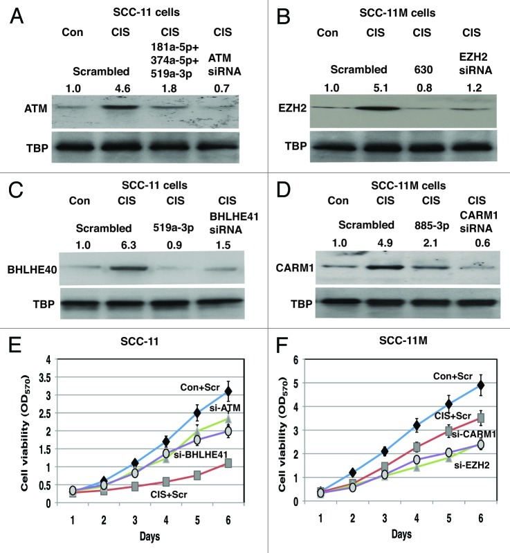
Figure 7. Specific microRNA mimics modulated expression of the Δ;Np63α protein interacting targets in SCC cells exposed to cisplatin and affected cell viability. Immunoblotting assay. SCC-11 cells (A and C) and SCC-11M cells (B and D) were transfected with the scrambled microRNA and indicated microRNA mimics for 36 h. Cells were then treated with control medium without cisplatin (Con) or medium with 10 μg/ml cisplatin (CIS) for additional 12 h and nuclear lysates were tested for indicated endogenous proteins. Loading levels were tested using a TBP antibody. Relative protein levels normalized for the TBP levels were quantified and shown above immunoblot images. Protein levels in cells with the scrambled miR were designated as 1. Cell viability assay. (E) SCC-11 cells were transfected with the scrambled siRNA (Scr) and siRNAs against ATM (si-ATM) or BHLHE41 (si-BHLHE41). (F) SCC-11M cells were transfected with the scrambled siRNA (Scr) and siRNAs against CARM1 (si-CARM1) or EZH2 (si-EZH2). Resulting cells were cultured in the presence (CIS) or absence (Con) of the 10 μg/ml cisplatin for indicated times. 104 cells/well in 96-well plates were then incubated in serum-free medium with 5 μg/ml of the 3-(4,5-dimethyl thiazol-2-yl)-2,5-diphenyl tetrazolium bromide in the dark for 4 h at 37°C. Cells were lysed and incubated for 2 h at 37°C, and the measurements were obtained on a Spectra Max-250 plate reader. Each assay was repeated at three times in triplicate. The bars are the mean ± SD of triplicate; p < 0.05, t-test.

An official website of the United States government
Here's how you know
Official websites use .gov
A
.gov website belongs to an official
government organization in the United States.
Secure .gov websites use HTTPS
A lock (
) or https:// means you've safely
connected to the .gov website. Share sensitive
information only on official, secure websites.
