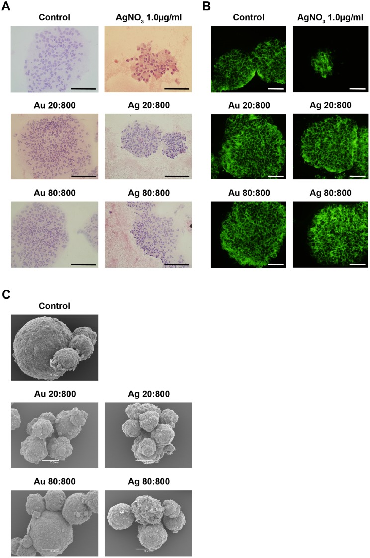
Figure 5. Gross morphology of HNPCs after Au- or AgNP exposure.
A. The neurospheres were stained with Htx-eosin after NP-exposure. The NPs affected the single cell morphology seen as changed orientation of the nuclei and cytoplasm. Spheres that were exposed to NPs had increased number of nucleus and they were more centred, while the cytoplasm was turned towards the sphere edge. The NP-exposure resulted the spheres more loosen, increased the number of pyknotic cells, and induced channel formation. Au 20∶800, gold 20 nm and 800 particles/cell; Au 80∶800, gold 80 nm and 800 particles/cell; Ag 20∶800, silver 20 nm and 800 particles/cell; Ag 80∶800, silver 80 nm and 800 particles/cell; AgNO3 1.0, silver nitrate 1.0 mg/ml. Scale bar equals 214 µm. B. The neurospheres were stained with Phalloidin after NP-exposure. NP exposure resulted in over-all altered cytoskeleton architecture, indicated by the increase in actin network. Au 20∶800, gold 20 nm and 800 particles/cell; Au 80∶800, gold 80 nm and 800 particles/cell; Ag 20∶800, silver 20 nm and 800 particles/cell; Ag 80∶800, silver 80 nm and 800 particles/cell; AgNO3 1.0, silver nitrate 1.0 mg/ml. Scale bar equals 50 µm. C. Scanning electron microscopic evaluation of the effects of AuNPs and AgNPs on HNPCs. NPs were added to the medium 48 h after seeding and spheres were prepared for scanning electron microscopy two weeks later. NP-exposure resulted in loose aggregates and fuzzy spheres compared to control spheres, which were smooth and compact. The morphological changes were more profound with increased concentration of the NPs. However, AgNPs affected the morphology more than AuNPs. Au 20∶800, gold 20 nm and 800 particles/cell; Au 80∶800, gold 80 nm and 800 particles/cell; Ag 20∶800, silver 20 nm and 800 particles/cell; Ag 80∶800, silver 80 nm and 800 particles/cell. Scale bars equal 50 µm.

