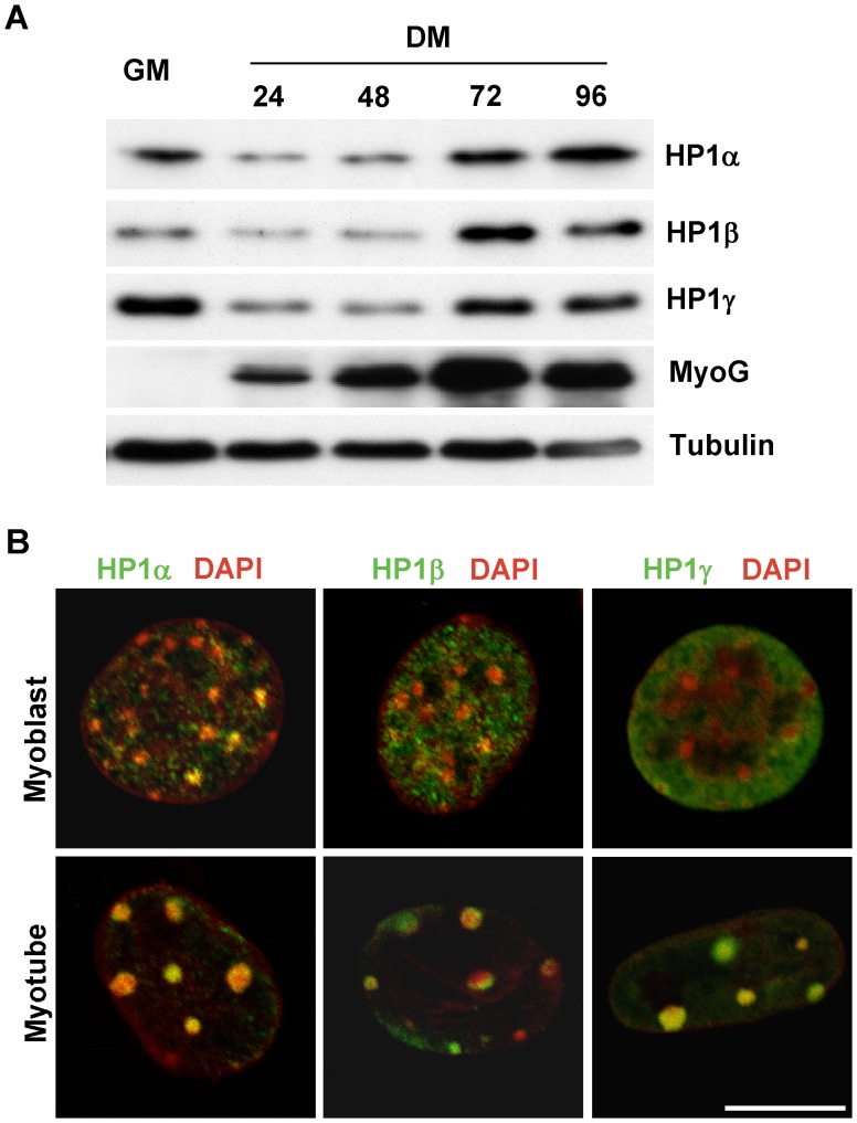
Figure 1. Expression and nuclear distribution of HP1 proteins during skeletal muscle differentiation.
A. C2C12 cells were cultured in GM (growth medium) or DM (differentiation medium) and collected at indicated time points. Total protein was extracted and subjected to Western blotting. B. Confocal fluorescence microscopy was performed on C2C12 myoblasts and myotubes after immunostaining for the indicated HP1 protein (green) and DAPI (converted to red). Scale bar equals 10 µm.

