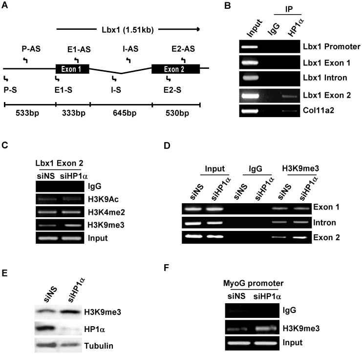
Figure 5. H3K9me3 levels on myogenic genes increased in C2C12 myoblasts after depleting HP1α.
A. Schematic diagram of the genomic structure of the mouse Lbx1 gene and locations of primers used for subsequent ChIP experiments. B. Protein-DNA complexes from cross-linked chromatin extracted from C2C12 myoblasts cultured in GM were immunoprecipitated with HP1α or mouse IgG. Bound DNA was amplified using the indicated PCR primers. C, D, C2C12 myoblasts were transfected with indicated siRNA, 48 hours after transfection, cross-linked chromatin was extracted and immunoprecipitated with indicted antibodies. Lbx1 exon 2 (C) or Lbx1 genomic sequences including exon 1, intron and exon 2 (D) were amplified. E. C2C12 myoblasts were transfected with the indicated siRNA, 48 hours after transfection total cell lysates were subjected to Western blotting with the indicated antibodies. F. C2C12 myoblasts were transfected with indicated siRNA, 48 hours after transfection cross-linked chromatin was extracted and immunoprecipitated with anti-H3K9me3 antibody. Precipitated DNA was used for PCR with primers spanning the MEF2-binding site on the myogenin gene promoter.

