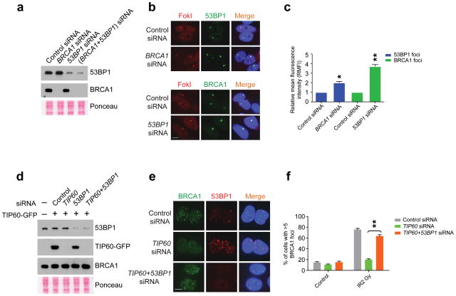
Figure 5. 53BP1 knockdown restores BRCA1 localization to DSBs.
(a) IB with cell lysates prepared from cells treated with siRNAs as indicated. (b) Representative images of 53BP1 (upper panel) or BRCA1 (lower panel) at mCherry-LacI-FokI-induced DSBs in cells transfected with the indicated siRNA. Scale bar, 10 μm. (c), Quantification of relative mean fluorescence intensity (RMFI) from b. RMFI was calculated from 200 cells and 3 independent experiments. Error bars represent S.E.M. * indicates p<0.05, ** p<0.005. (d), IB with cell lysates prepared from TIP60-GFP expressing cells treated with siRNAs as indicated. (e) Representative images of BRCA1 and 53BP1 co-staining following fixation at 5 hours after 2 Gy IR treatment of U2OS cells that were transfected with the indicated siRNA. Scale bar, 10 μm. (f) Quantification of percentage of cells possessing greater than 5 BRCA1 IRIF per cell from e. Error bars represent S.E.M. ** indicates p<0.005.

