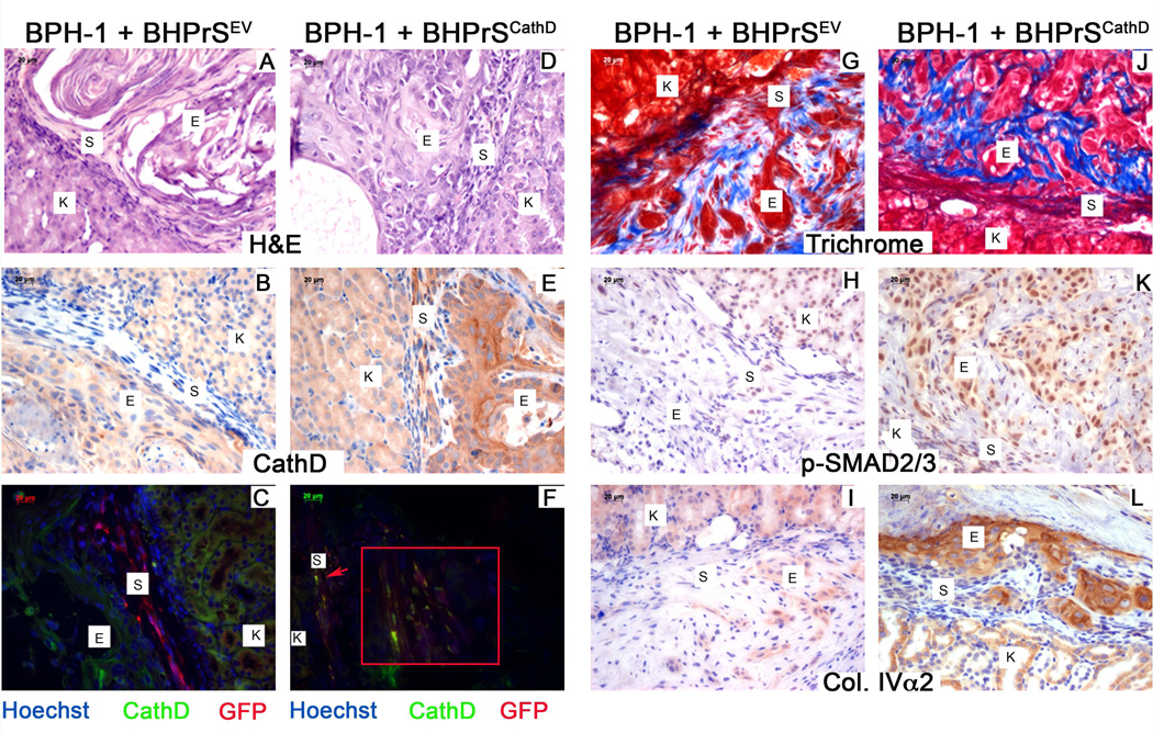
Figure 5. Overexpression of Cathepsin D induces a malignant transformation through activation of TGFβ signaling.
(A) Characterization of CathD overexpressing grafts. H&E staining (top) of BPH-1 + BHPrSEV and BPH-1 + BHPrSCathD. Recombinations of BPH-1 + BHPrSCathD, produced malignant transformations. IHC for CathD (middle) strong expression visible in the stroma of recombinations of BPH-1 + BHPrSCathD. Immunoflurescence for CathD (green) and GFP (red) show co-localization for GFP and CathD (yellow) in recombinations of BPH-1 + BHPrSCathD. (B) Masson’s trichrome staining (top) of recombinations of BPH-1 + BHPrSEV and BPH-1 + BHPrSCathD. IHC for p-SMAD2/3 (middle) and Collagen IV±2 (lower) in recombinations of BPH-1 + BHPrSEV and BPH-1 + BHPrSCathD. Scale bar equal to 20µm.

