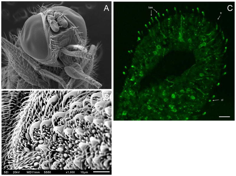
Figure 3.
Localization of HirrOrco in H. irritans male antenna. Green fluorescence represents anti-Orco detected using an Alexa Fluor 488-conjugated secondary antibody. (A) SEM of a horn fly head depicting the three antennal (ant) segments (I: scape, II: pedicel, III: flagellum), the maxillary palp (mp), and the proboscis (prob). (B) A region of the flagellum identifying trichoid, basiconic, and clavate sensilla, as described by White and Bay (1980). (C) HirrOrco labeling of nerve cell bodies and dendrites within specific antennal sensilla. Different sensillar types were labeled by anti-Orco, namely basiconic (bas), clavate (cl) and trichoid (tr) sensilla. Scale bar represents 20μm.

