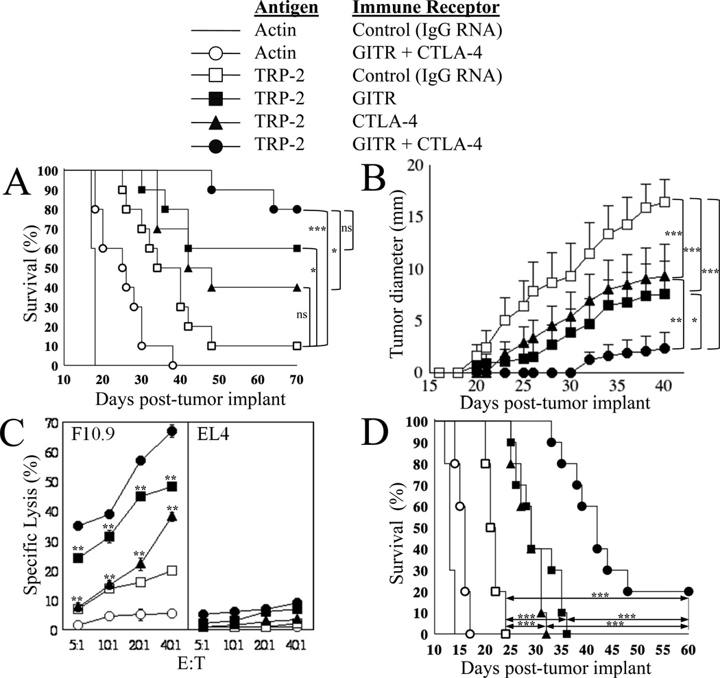
Figure 1. Effect of local immune receptor modulation on anti-tumor immunity in response to tumor-specific immunization in a B16/F10.9 melanoma immunotherapy model.
(A) 7–8-week old C57BL/6 mice (10/group) were implanted with 2.5x10⁴ B16/F10.9 tumor cells s.c. in the flank region, then vaccinated 2 days later. The Figure key depicts the antigen used and the immune receptor targeted in each group. For experiments testing the combination of TRP-2 tumor antigen mRNA-transfected DCs and mAb-encoding mRNA-transfected DCs, mice were immunized s.c. at the base of each ear pinna with 1x10⁵ DCs for each DC group in 50 µl PBS/ear pinna. Total number of DCs per mouse was 4x10⁵ for groups testing 2 combinations and 6x10⁵ DCs per mouse for groups testing 3 combinations. Starting on day 10, tumor growth was monitored every other day by measuring the appearance of palpable (5–6 mm diameter) tumors and mice were sacrificed once the tumor diameter reached 20 mm. All surviving mice were tumor-free at day 120 with no evidence of autoimmunity, at which time they were sacrificed. Comparisons between groups at day 70 were performed using the log-rank test (Mantel-Haenszel test), *p<0.05, ***p≤0.001, ns=non-significant.
Median survival day: TRP-2+IgG: 37; TRP-2+anti-CTLA-4: 45; actin+anti-GITR+anti-CTLA-4: 25.5 and actin+IgG: 18. All other groups, median: undefined.
(B) In these same groups of mice, average tumor size ± SEM versus days post-tumor implant is displayed. Tumor growth curves over time were compared using one-way ANOVA for repeated measures (p<0.0001) with Bonferroni multiple comparison post-test to compare the four groups indicated, *p≤0.05, **p≤0.01, ***p≤0.001.
(C) 7–8-week old C57BL/6 mice (2/group) were implanted with 2.5x10⁴ B16/F10.9 tumor and immunized as indicated in (A). Cells were harvested from the draining auricular lymph nodes 10-days post-immunization and the non-adherent cells were stimulated with DCs transfected with TRP-2 mRNA. Induction of TRP-2-specific CTLs was determined 5-days post-restimulation using a standard cytotoxicity assay as described in Materials and Methods. F10.9 and EL4 cells were used as targets. The standard deviation reflects variation between triplicate wells. ** p<0.01 as compared to TRP-2+anti-GITR+anti-CTLA-4 group (filled circles) using paired two-tailed Student’s t test.
(D) 7–8-week old C57BL/6 mice (10/group) were implanted with 3.0x10⁴ B16/F10.9 tumor cells s.c. in the flank region. Mice were vaccinated once 7-days after tumor implantation, at which time 3mm diameter s.c. tumor deposits were palpable in all mice. DCs were transfected with the mRNA combinations as indicated in the Figure key. Mice were immunized s.c. at the base of each ear pinna with 1.5x10⁵ DCs in 50 µl PBS/ear pinna. Total number of DCs per mouse was 3x10⁵ for all groups tested. Starting on day 8, tumor growth was monitored and recorded when tumor size was 5mm and above. Mice were sacrificed once the tumor diameter reached 20 mm (used to determine percent survival). All surviving mice were tumor-free at day 60 with no evidence of autoimmunity, at which time they were sacrificed. Comparisons between groups on day 50 were performed using the log-rank test (Mantel-Haenszel test), ***p≤0.0001.
Median survival day: TRP-2+IgG: 21.5; TRP-2+GITR-L: 29; TRP-2+anti-CTLA-4: 29; TRP-2+GITR-L+anti-CTLA-4: 42; actin+GITR-L+anti-CTLA-4: 16 and actin+IgG: 13.

